Product Features
Full digital imaging technology
Crystal-clear Image
Computer platforms Abundant functions
Powerful image and report management
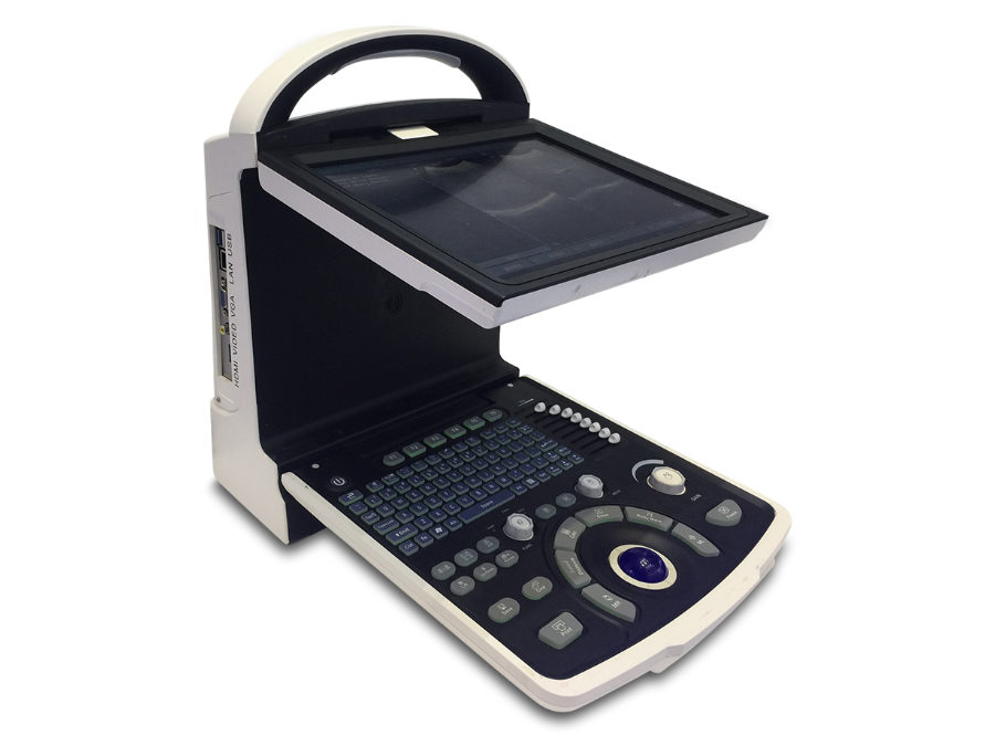
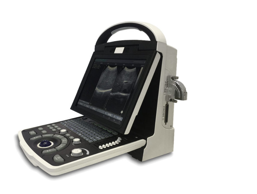
10 inch no interlaced CRT monitor
Silica gel backlight keyboard
DICOM display and transfer
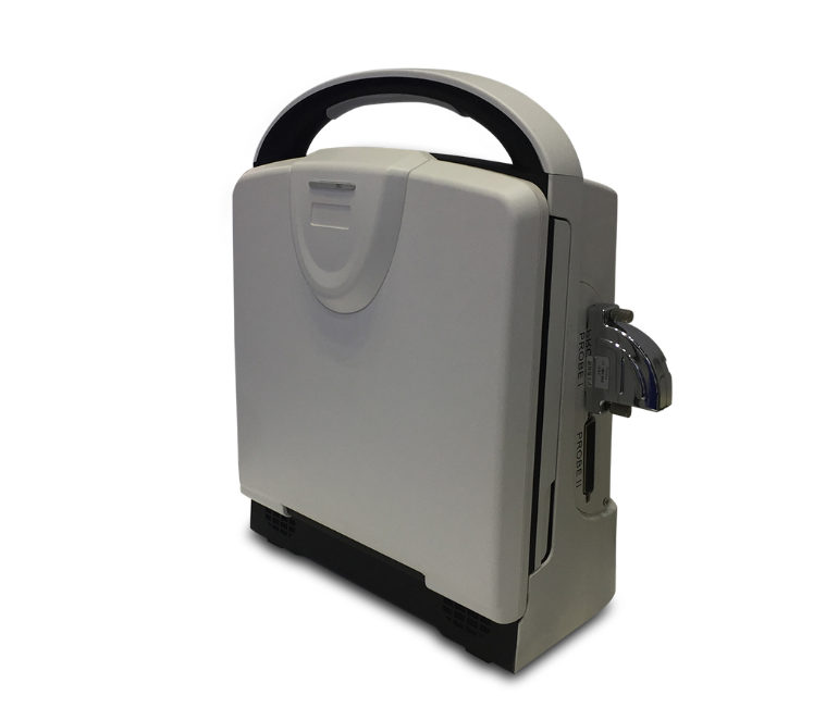
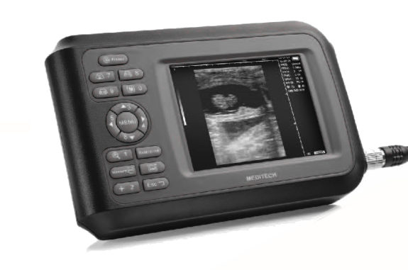
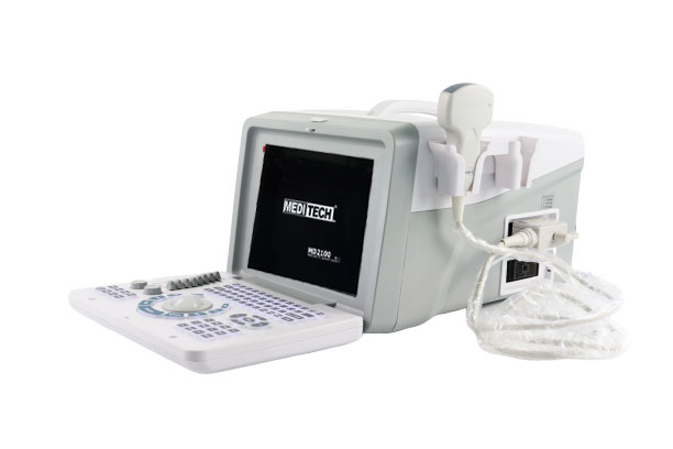
Pretty portable and light
Large volume Storage and cine loop
128/256/512/1024 frame cine loop
Broadband Multi frequency probes
8 segment TGC 16 step zoom
Complete application software packages
2 probe connector
Compatible with USB laser/inkjet printers
3D Image Function(optional)
-
THI
-
Imaging technologies
-
Intelligent Speckle Reduction
-
3D reconstruction
-
Abundant user management functions
-
One key recording
-
User defined OB table
-
Expert vocabulary and report templates
Product Specifications
Displaying mode: B mode B/B mode B/M mode M mode 4B mode
Scanning mode: Electric convex Electric linear Electric micro-convex
Displaying depth: 20mm-310mm
Probe port: 1 OR 2
Voltage: AC100V-240V
Output port: USBDICOMVGA
Gray scale: 256
Time gain: Gain:10~255dB adjustable ;time gain compensation(TGC): 8 segments which can be adjusted independently in B mode and M mode.
Storage function: image storage cine loop storage mass storage capacity≥10G.measurement result storage report storage
Cine-loop: 641282565121024 frames can be chosen;auto/manual cine loop
Patient information: Hospital name; patient name; case number; patient age; gender;
Focusing mode: 1-4 selectable focus multiple combination way focus position moving available
Real-time zoom local zoom: Multi-stage display rate(depth ascension)function roll line function
Annotation function: Annotation at the image area
Annotation of patient information
Body mark: 140
Cursor: The combination of Trackball and keyboard
Color display: 43 color display function
DC-AC: AC/DC
Report: Display storage and print report directly.
Variable angle scanning: 16kinds of angle scanning include:25%30%35%…100
Image processing: Pre-processing post-processing dynamic range frame correlation line correlation edge enhancement black/white conversion left/right conversion up/down conversion contrast brightness gamma correction
Dynamic range: Continuously adjustable: 20~150dB
Measurement and calculation: B mode: distance circumstance area volume angle histogram hip joint angle
M mode: heart rate time slope
Abdomen: uterus cervix endometrial ovary ovarian follicle cyst enclosed mass
Obstetric: 14(ACBPDCRLFLTHDGSOFDHUMERUSTIDIAULNAHCTADAPTDFTA)measure gestation age fetal weight AFI
Cardiology: LV.LV function LVPWRVAWT aorta mistral valve
Urology: L/R kidney volume bladder volume prostate volume
Small organs: Ophthalmology. Thyroid residual urine volume
Related Accessories
Standard:
Multi frequency convex probe ...... 1Pcs
Power Cable ............................... 1Pcs
Cleaning Cloth ............................ 1Pcs
Options:
7.5MHz high-frequency linear array probe
6.5MHz intra-cavity (trans-vaginal) probe
7.5MHz rectal probe
3.5MHz micro-convex probe
B/W Laser printer
Footswitch
Battery
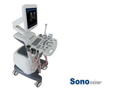
Trolley
Ultrasonic work-station
Carrying Bag